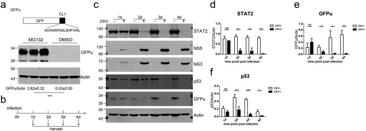
Figure 3.
ZIKV infection accelerated proteasomal degradation. (a) Huh7.5 cells stably expressing GFPu (upper panel) in triplicate wells were treated with DMSO or MG132 (10 μM) for 24 h and then analysed by western blotting with the indicated antibodies. (b) Schematic of the experimental design for c–i. Huh7.5-GFPu cells were infected (+) or not (−) with ZIKV C7 (MOI = 1) and harvested at the indicated time points. (c) Western blotting analysis of the cells with the indicated antibodies. Representative pictures of three biological replicates are shown. The values to the left of the blots are molecular sizes in kilodaltons. (d–f) The protein abundances of each protein in B were quantified and plotted. The mean ± SD of three biological replicates is shown (n = 3). Statistical analysis was performed between C7-infected groups and uninfected groups (ns, not significant, *P < 0.05, **P < 0.01, ***P < 0.001; two-tailed, unpaired t-test).

