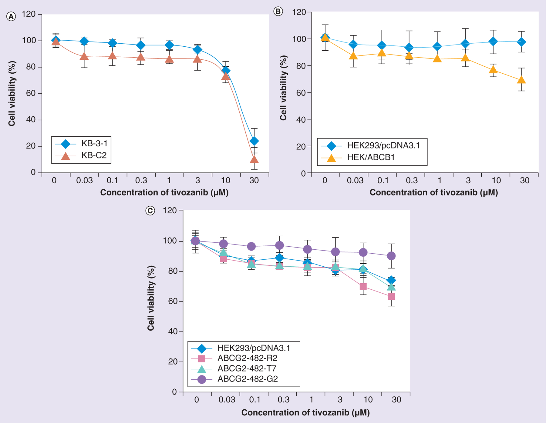
Figure 1. Cytotoxicity of tivozanib.

(A) Cytotoxicity of tivozanib in KB-3-1 and KB-C2 cell lines. (B) Cytotoxicity of tivozanib in HEK293/pcDNA3.1 and HEK/ABCB1 cell lines. (C) Cell cytotoxicity of tivozanib in HEK293/pcDNA3.1, ABCG2-482-R2, ABCG2-482-T7 and ABCG2-482-G2 cell lines. The data points represent the mean ± standard deviation of triplicate determinations.
