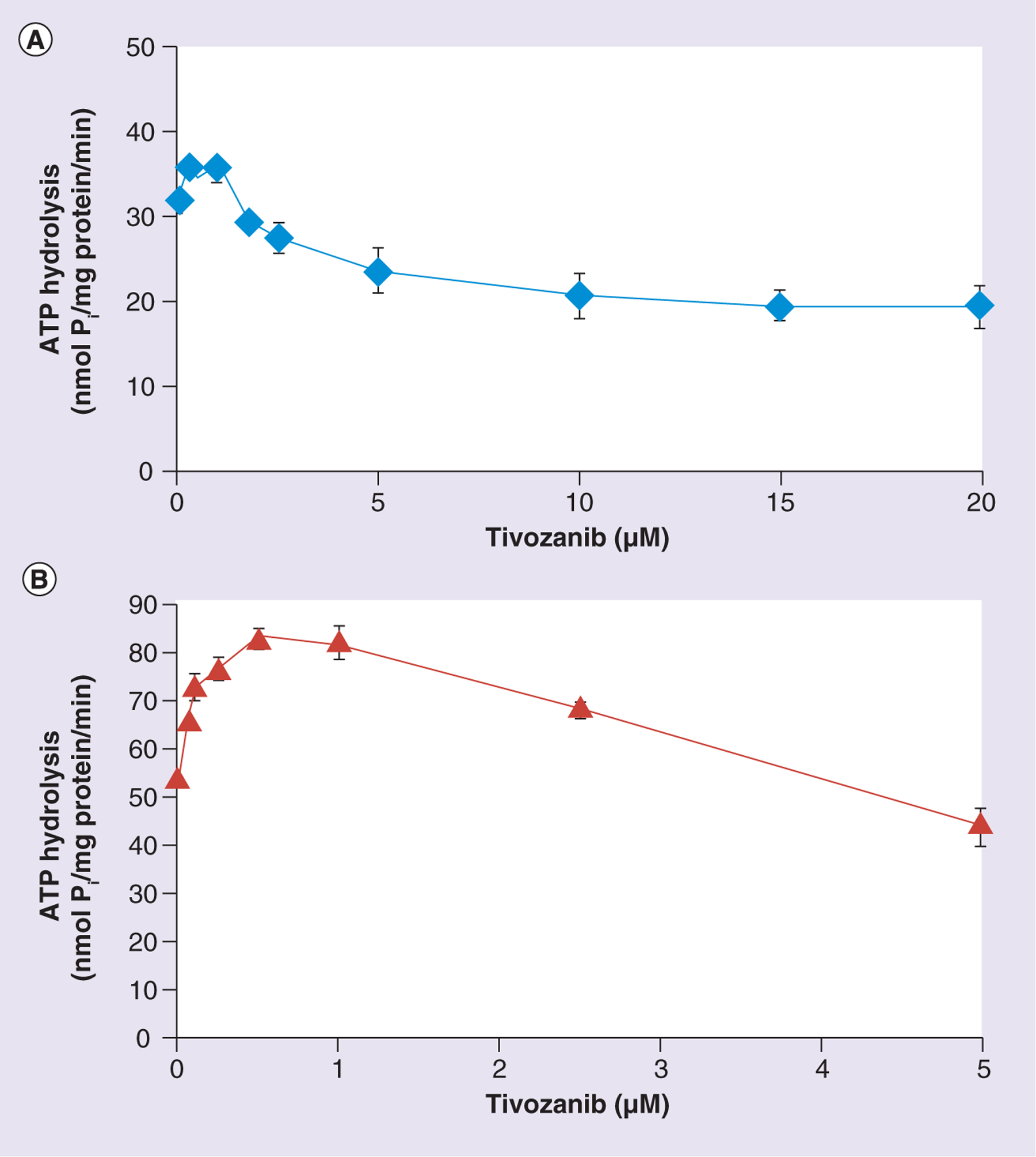
Figure 6. Modulation of the basal ATPase activity of ABCB1 and ABCG2 by tivozanib.

Crude membranes (10 μg protein/100 μl) from High Five™ (Life Technologies, NY, USA) insect cells expressing (A) ABCB1 or (B) ABCG2 were incubated with increasing concentrations of tivozanib (0–20 μM) in the presence or absence of sodium orthovanadate (Vi) (0.3 mM) in an ATPase assay buffer, as described in the ‘Materials & methods’ section. The reaction was initiated by the addition of 5 mM ATP, incubated for 20 min at 37°C and then terminated by the addition of 5% sodium dodecyl sulfate solution. The amount of Pi released was quantified using a colorimetric reaction. The data points represent the means from at least three independent experiments, while the bars represent the standard deviation values.
Pi: Inorganic phosphate.
