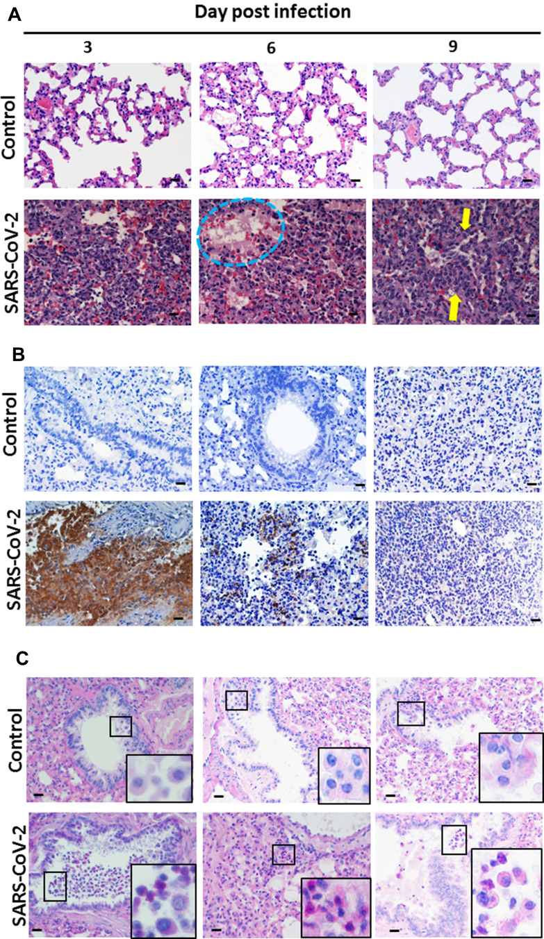
Figure 4.
Pathological changes in SARS-CoV-2-infected hamster lung on days 3, 6, and 9 post-infection. Syrian hamsters aged 6 to 8 weeks (n=5 for SARS-CoV-2, and n=3 for non-infected control) were infected with 5×10⁵ TCID50 of SARS-CoV-2 on day 0. On days 3, 6, and 9, samples of lung and trachea tissue were removed, fixed in formalin and embedded in paraffin using routine methods, and then processed for staining. (A) Samples of lung sections stained with H&E. The blue dotted circle outlines hyaline membrane formation. The yellow arrows present macrophage aggregates in the airway and alveolar spaces. (B) Lung sections immunostained with anti-SARS-CoV nucleoprotein antibody. (C) PAS staining showing mucus expression in hamster lung. Small box frames show macrophages that scavenged the extra mucus near the trachea. Large box frames show enlargements of macrophage phagocytosis. Scale bar for the 400× panels = 20 µm.

