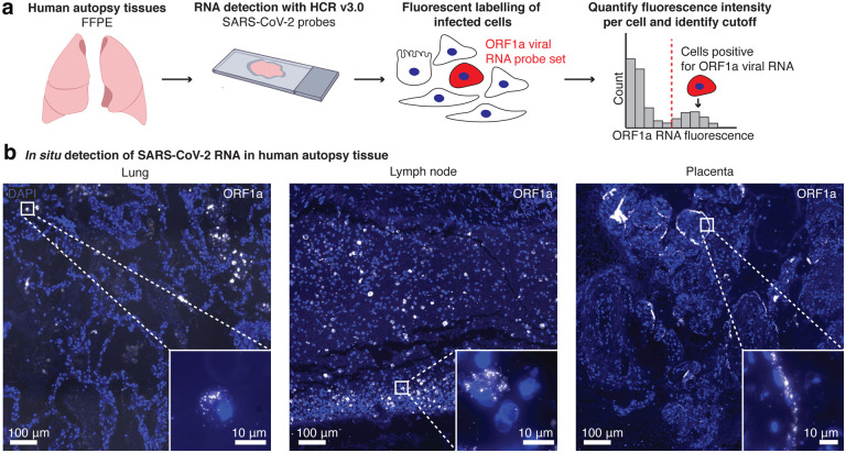
Figure 2: RNA FISH HCR in FFPE human autopsy tissues.
a. Experiment design in which we performed RNA FISH HCR with ORF1a probe sets on FFPE tissues including lung, hilar lymph node, and placenta. b. Example images of each tissue with ORF1a RNA staining. Images are large area scans of image tiles acquired at 20X. Scale bar on the large images shows 100 μm. Inset images show a zoomed in example of ORF1a RNA staining in that tissue. Scale bars on these inset images are 10 μm. DAPI stain (blue) labels the cell nuclei in all images.

