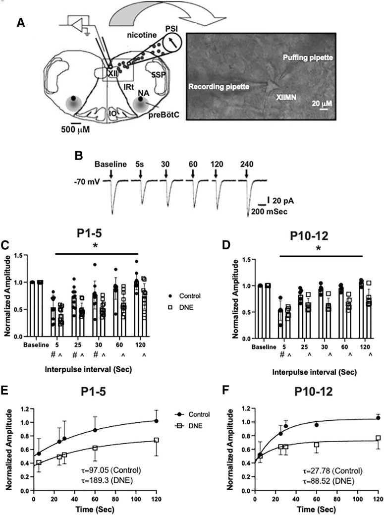
Figure 7.
nAChR desensitization and recovery. A, A representation of the experimental set-up showing the rostral surface of the medullary slice preparation with landmarks including the XII motor nucleus, trigeminal nucleus (5SP), preBötzinger complex, nucleus ambiguus (NA), inferior olivary nucleus (IO), and the intermediate reticular formation (IRt). Inset shows a high-resolution image (40×) of an isolated XII MN with a recording pipette and a “puffing” pipette. B, Examples of inward currents produced by nicotine pressure pulses (2 mm; 30 ms) in a P4 conditioning neuron during a 4-min pressure-pulse protocol. Pressure pulses occurred at successively longer intervals following a conditioning pulse at time 0. At both P1–P5 (C) and P10–P12 (D), eDNE (open squares) causes a delay in the recovery of nAChR-mediated peak currents compared with control (closed circles). E, F, Quantification of nAChR recovery using a single exponential fit to the average data shown in C, D. eDNE slows recovery at both age groups as indicated by time constants (τ), shown in insets. Data derived from n = 12 control neurons and n = 12 eDNE neurons in each age group. All data presented as mean ± SD; in C, D # indicates significant difference from baseline within control group; ^ indicates significant difference from baseline within eDNE group. A reproduced with permission from Pilarski et al. (2012).

