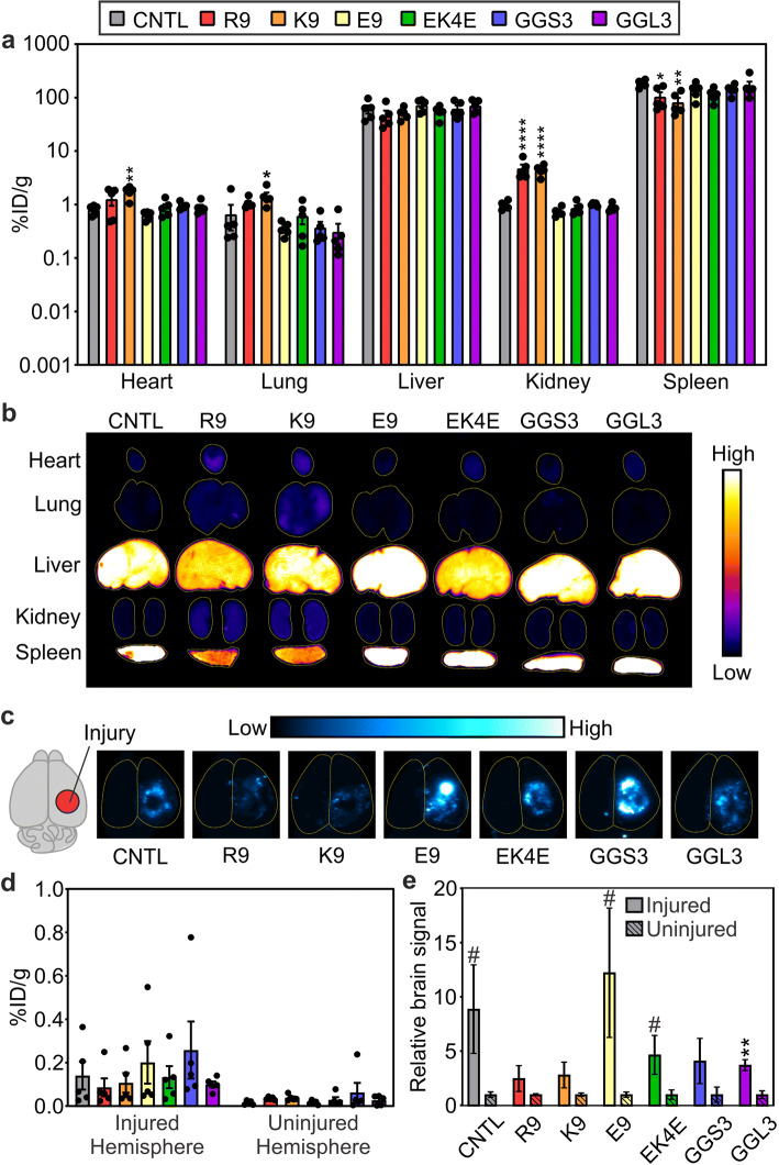
Figure 5.
Accumulation of peptide-modified nanoparticles in dissociated organs 1 h after administration (a) and representative surface fluorescent images (n = 5, mean ± SEM; one-way ANOVA with Bonferroni post-test compared to control nanoparticles, *p < 0.05, **p < 0.01, ****p < 0.0001) (b). Representative surface fluorescent images (c) and accumulation of peptide-modified nanoparticles in dissociated brain tissue, separated by injured and contralateral hemispheres, 1 h after administration (d). Relative amounts of nanoparticle signal in the injured vs. contralateral uninjured hemisphere (n = 5, mean ± SEM; two-tailed t test between injured and uninjured groups, #p < 0.1, **p < 0.01) (e)

