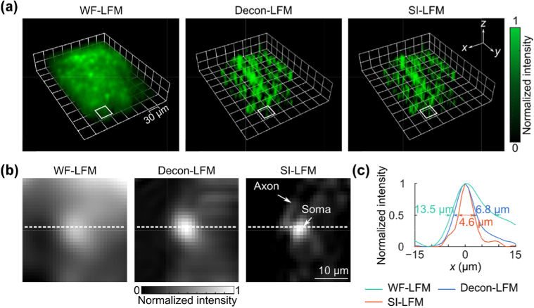
Fig. 4.
– SI-LFM resolved the sub-cellular structure of neurons labeled by GCaMP6f within a fixed mouse retina with more detail than WF-LFM. (a) Volumetric images showed that SI-LFM acquired sharp retina features missed by the other two imaging modalities. All three images are thresholded at a normalized intensity of 0.4 such that only values above this threshold are shown. Individual cells appear as refined footprints in the SI-LFM volumetric rendering, but lose resolution in the WF-LFM and Decon-LFM images. (b) The magnified views of the white boxes in Fig. 4(a) show the soma and axon of a neuron in the SI-LFM image, but not in images acquired by other modalities. (c) The intensity profile along the dashed line in panel (b) showed that among the three light-field imaging modalities, SI-LFM imaged the cell with the smallest FWHMs along the x-direction.

