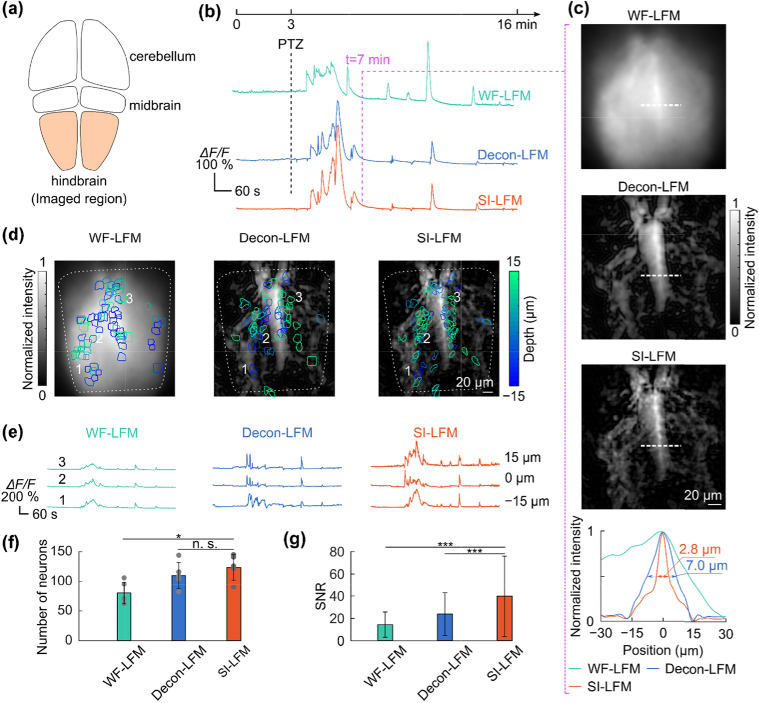
Fig. 5.
– SI-LFM detected active neurons in imaging living zebrafish larvae with high fidelity. (a) The schematic illustration shows sub-regions of the larval zebrafish brain, including the hindbrain target. (b) PTZ delivered at 3 min (top) induced seizures and large calcium transients in zebrafish (bottom). (c) At 7 min, the image acquired with SI-LFM at 2 µm depth resolved more brain structures than images acquired with the other two imaging modalities. The intensity distribution of the features labeled along white dash lines (bottom) was sharper. (d) The depth-coded colored masks labeled the active neurons imaged with different imaging modalities. (e) The calcium activity of representative neurons located at different depths indicated that SI-LFM recorded the same neural activity with the highest change in fluorescence. (f) SI-LFM identified significantly more active neurons than the WF-LFM (dots are individual data values; *p < 0.05, n.s. – not significant, two-sided Wilcoxon rank-sum test, n = 5 fish; error bars are standard deviations). (g) The calcium transients recorded by SI-LFM had significantly higher SNR than the transients recorded by other LFM techniques (***p < 0.001, *p < 0.05, n.s. – not significant, two-sided Wilcoxon rank-sum test; n = 625, 555, and 405 neurons respectively from SI-LFM, Decon-LFM, and WF-LFM, all from 5 fish; error bars are standard deviations).

