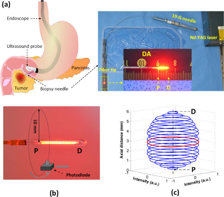
Fig. 1.
Circumferential interstitial laser ablation (CILA) of pancreatic tissue: (a) illustration of endoscopic ultrasound (EUS)-guided CILA on pancreatic adenocarcinoma with diffusing applicator through small biopsy needle (19-G), (b) uniform HeNe light distribution along diffusing applicator (P = proximal and D = distal ends), and (c) 3D normalized spatial emission profile measured by photodiode in (b). Red color represents the polar intensity measured 3 mm away from P (normalized polar intensity = 0.9 ± 0.1).

