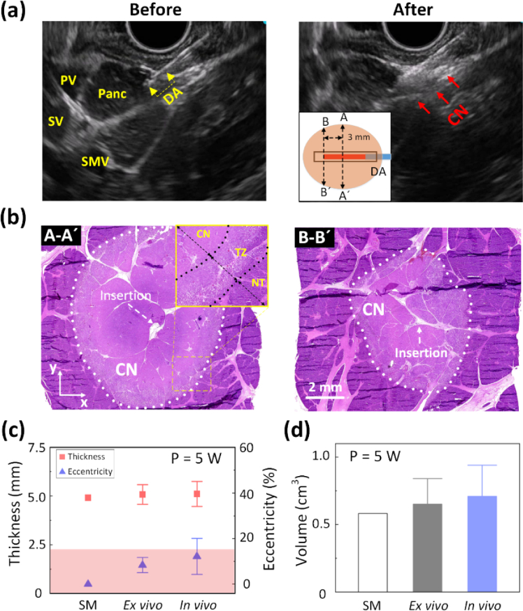
Fig. 6.
EUS-guided CILA on pancreatic tissue in in vivo porcine models (5 W for 200 s; 1000 J delivered): (a) US images captured before (left) and after (right) CILA (DA = diffusing applicator; PV = Portal vein; SV = Splenic vein; SMV = Superior mesenteric vein; Panc = Pancreas; CN = coagulation necrosis), (b) histology images (40) of transverse tissue sections (xy plane) from A-Á(middle of DA; left) and B-B´ (3 mm away from distal end of DA; right), and (c) quantitative comparisons of ablation thickness and eccentricity (left) and ablation volume (right) among simulation (SM), ex vivo tests (N = 10), and in vivo tests (N = 2), and radiofrequency ablation (RFA). An inlet in (a) illustrates coagulation necrosis around DA in the tissue. An inlet in (b) presents microscopic changes in the ablated tissue (100; TZ = transition zone; NT = native tissue).

