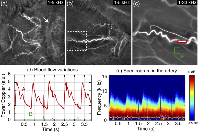
Fig. 1.
Blood flow imaging in episcleral and conjunctival vessels. (a-c) Power Doppler images, the arrow points to the junction between the conjunctiva and the iris. (d) Power Doppler variations in the indicated ROIs. (e) Spectrogram measured in the artery. Visualization 1 (25.5MB, mp4) .

