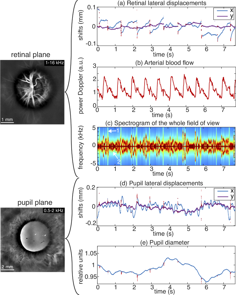
Fig. 4.
Tracking the movements of an eye slightly affected strabismus with LDH. In the retinal plane are measured (a) the fundus lateral displacements, (b) the retinal arterial blood flow, and (c) the averaged Doppler spectrogram of the fundus revealing the signatures of in-plane saccades and out-of-plane motion. In the anterior segment reconstruction are tracked (d) the pupil lateral displacements, and (e) the pupil diameter. Visualization 5 (29.6MB, mp4) shows the 6-16 kHz power Doppler movies in the retinal and pupil planes.

