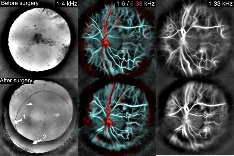
Fig. 6.
Imaging an eye affected by cataract before and after surgery. Before the operation, opacities are present in the center and periphery of the eye lens, but fundus imaging with LDH can still be performed. After the operation, the mark of the implant is visible in the anterior segment, and a similar quality of imaging is obtained in the fundus.

