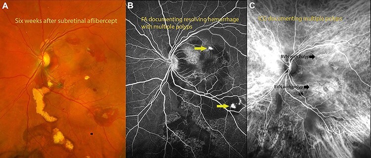
Figure 3 .

(A) fundus photograph 6 weeks after surgery documenting resolving hemorrhage with residual exudates from polyps. (B) Flourescein angiography documenting polyps with residual hemorrhage. (C) Indocyanin angiography demonstrating areas of polyps with BVNs in the areas of polyps.
