
Figure 1.
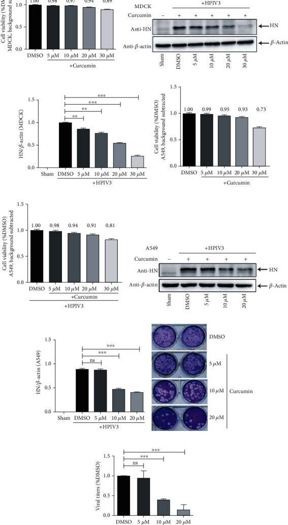
Curcumin inhibits HPIV3 infection. (a) MDCK cells were incubated with different concentrations of curcumin (5 μM, 10 μM, 20 μM, and 30 μM) or control DMSO for 48 h; the cell viability was examined by the CCK-8 assay as per the manufacturer's instruction. There was no significant toxicity when cells were treated with 5 μM, 10 μM, and 20 μM curcumin. (b, c) MDCK cells were treated with the abovementioned concentrations of curcumin or DMSO for 6 h; then, cells were washed and infected by HPIV3 (MOI = 0.1) with different concentrations of curcumin present. The sham group was neither virus-infected nor drug-treated. Cells were collected after 36 h infection, viral HN protein was analyzed by western blot (WB), and cellular β-actin was used as a loading control. HN/β-actin for each group was calculated as described in Materials and Methods. Data are means ± SD from three experiments. Student's test: ∗∗p < 0.01 and ∗∗∗p < 0.001. A549 cells noninfected (d) or infected with HPIV3 (e) were incubated with different concentrations of curcumin (5 μM, 10 μM, 20 μM, and 30 μM) or control DMSO for 48 h; the cell viability was examined as above. (f, g) A549 cells were treated with curcumin and infected with HPIV3 as described in (b, c). Cells were collected after 36 h infection, viral HN protein and cellular β-actin were analyzed by WB, and HN/β-actin for each group was calculated as above. Data are means ± SD from three experiments. Student's test: ns: nonsignificant; ∗∗∗p < 0.001. (h, i) Viral plaques and titers in the culture medium of (f, g) were determined by plaque assay as described in Materials and Methods. Data are means ± SD from three experiments. Student's test: ns: nonsignificant; ∗∗∗p < 0.001.