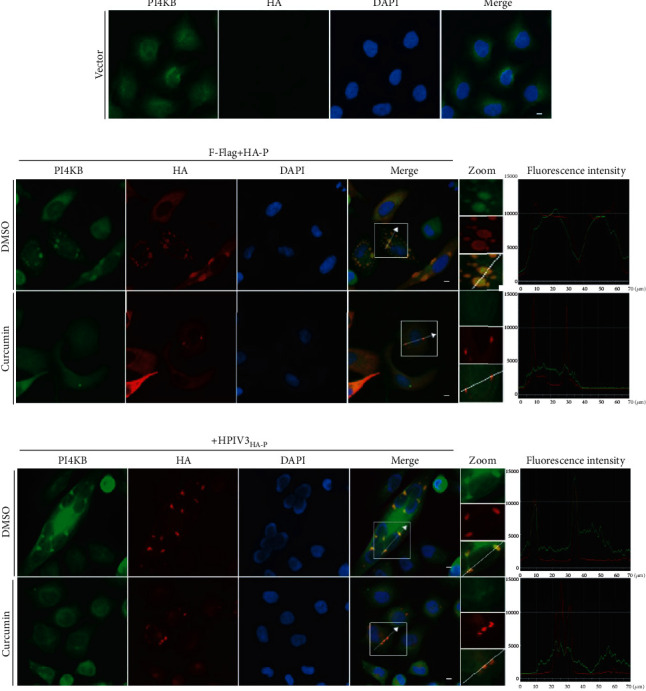
Figure 6.

Curcumin interferes with colocalization of PI4KB in IBs. (a) HeLa cells were fixed, and cellular PI4KB was stained with rabbit anti-PI4KB primary antibody and goat anti-rabbit AF488 as described in Materials and Methods. Scale bar = 10 μm. (b) HeLa cells were cotransfected with plasmids encoding N-Flag and HA-P jointly by lipo3000. DMSO or curcumin was added at 12 h before sample collection. At 24 h posttransfection, Cells were collected and fixed, PI4KB was stained as described above, HA-P was immunostained to visualize IBs, and nuclei were counterstained with DAPI. Scale bar = 10 μm. The fluorescence intensity profile of IBs (red) and PI4KB (green) was measured along the line drawn on a zoom panel by NIS-Elements BR 4.60.00.64-bit. (c) HeLa cells were infected with HPIV3HA-P at an MOI of 1.0; DMSO or curcumin was added at 12 h before sample collection. The cells were collected after 36 h infection. PI4KB and IBs were immunostained as described above, nuclei were counterstained with DAPI. Scale bar = 10 μm. The fluorescence intensity profile of IBs (red) and PI4KB (green) was measured along the line drawn on a zoom panel by NIS-Elements BR 4.60.00.64-bit.
