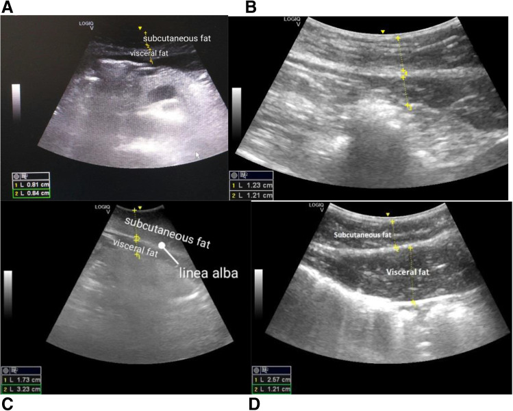
Fig. 4.
Ultrasound imaging as the diagnostic tool to discriminates between A abnormally low and B excessive versus C normal abdominal and visceral fat distribution and D SAT patterns in visceral fat redistribution; evident gender difference is well respected by gender-specific patterns of fat distribution, namely for males (B and D) and females (A and C). Notably, specific movement patterns at breathing further contribute to the correctness of the fat tissue measurement in abdominal cavity. The scanning was performed in the sagittal plane along the linea alba; the figure is adapted from [4]

