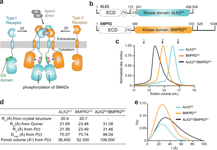
Fig. 1. The BMPR2 and ALK2 kinase domains form a thermodynamically stable heterodimeric complex in solution.
a Schematic diagram of the ligand-induced type I/type II receptor tetramer and resulting regulatory Gly/Ser rich (GS) domain phosphorylation. Pink dots symbolize phosphorylated residues within the GS domain. b Diagram depicting the domain boundaries within the ALK2 and BMPR2 receptors, including the extracellular domain (ECD), transmembrane (TM) helix, the GS domain, and the kinase domains (KD). Residues marking kinase domain boundaries and boundaries of the constructs used in this study are marked. c Size-exclusion chromatograms of ALK2KD (cyan), BMPR2KD (orange) and the ALK2KD/BMPR2KD complex (black) resolved on a Superdex 200 Increase 10/300 GL column. Molecular weight standards are indicated above: 1— γ-globulin (158,000 Da), 2—ovalbumin (44,000 Da) and 3—myoglobin (17,000 Da). d The radius of gyration (Rg) calculated from the crystal structures of ALK2 (PDB ID: 3MTF) and BMPR2 (PDB ID: 3G2F) compared with molecular dimensions calculated from the SEC-SAXS profiles of the ALK2KD, BMPR2KD, and the ALK2KD/BMPR2KD complex. e SAXS P(r) function of ALK2KD (cyan), BMPR2KD (orange), and the ALK2KD/BMPR2KD (black) complex.

