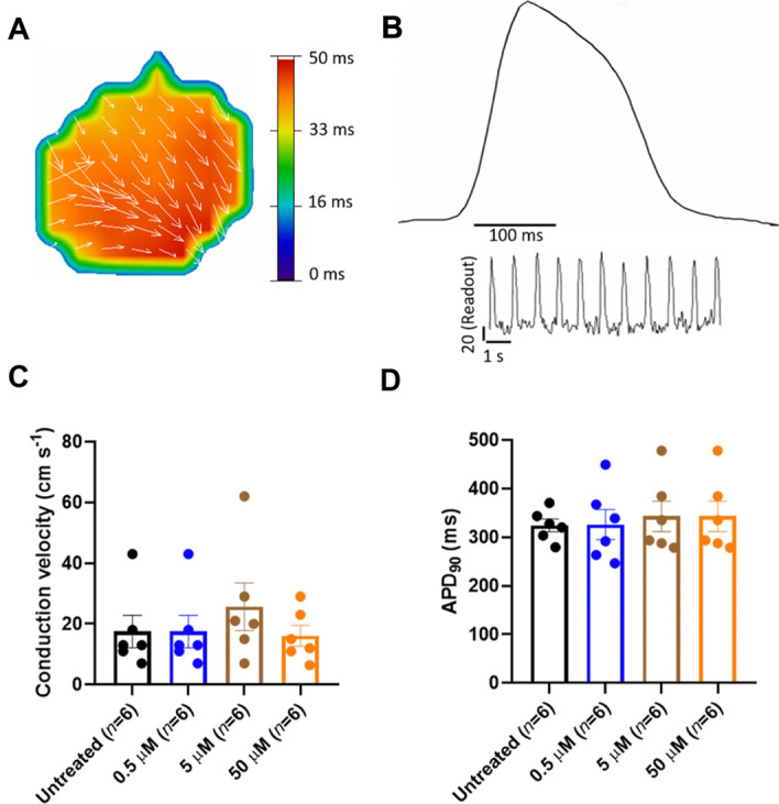
Figure 7.

Conduction velocity and APD90 after treatment of human iPSC-derived cardiomyocytes with the SERCA activator CDN1163. (A) Representative impulse propagation during the spontaneous activity of untreated monolayer syncytium formed of human iPSC-derived cardiac cells. Impulse propagation is represented by the activation maps of action potential propagation at different activation times, with the arrows showing the direction of the propagation wave across the monolayer syncytium. (B) Representative trace of the spontaneous action potential (AP) obtained by optical mapping of a syncytium formed by human iPSC-derived cardiomyocytes. The inset represents single-pixel signals of spontaneous optical AP recordings. (C) Conduction velocity (CV) calculated from each activation map in the presence and absence (untreated control) of CDN1163. We found no significant changes in CV between the control and treatment groups. (D) Quantification of APD90 from optical mapping readings shown in panel (B). We found that treatment of human iPSC-derived cardiomyocytes with CDN1163 does not have significant effects on the APD90 compared to the untreated control. Data are reported as mean ± SEM (n = 6). Groups were compared using a one-way analysis of variance (ANOVA) with the Dunnett test; statistical significance was set at p < 0.05.
