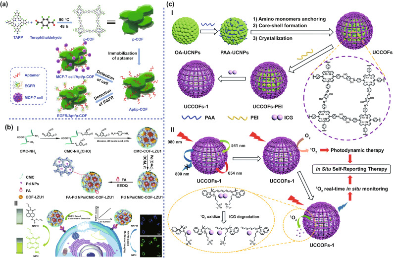
Fig. 7.
COFs for imaging and diagnosis. a Schematic diagram of the p-COF-based aptasensor for detecting EGFR or MCF-7 cells, including (1) preparation of p-COF, (2) immobilization of the aptamer strands, and (3) detection of EGFR or MCF-7 cells. Adapted with permission [106]. Copyright ©2018 Elsevier B.V. All rights reserved. b I. Illustration of the synthetic process of FA-Pd NPs/CMC-COF-LZU1. II. Schematic illustration of the dual-function FA-Pd NPs/CMC-COF-LZU1 for cancer cell imaging. Adapted with permission [107]. Copyright © The Royal Society of Chemistry 2020. c I. A schematic illustration of the design and synthesis of the upconverting COF nanoplatform UCCOFs-1. II. A schematic illustration of the NIR-excited in situ self-reporting PDT process. Adapted with permission [37]. Copyright © The Royal Society of Chemistry 2020

