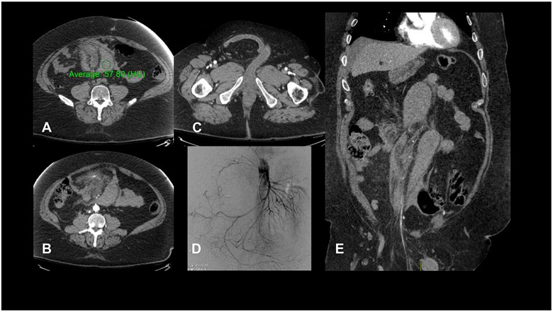
Figure:
CT imaging for the presented patient at the time of his ED visit. (A) High attenuation (57 HU on non-contrast imaging) crescentic mesenteric collection, consistent with mesenteric hematoma; (B) blush from a branch of the superior mesenteric artery, indicating the site of active bleeding; (C) right inguinal hernia with unremarkable small bowel; (D) conventional angiography demonstrating subtle beading of small superior mesenteric artery branches; (E) coronal depiction of two large hematomas and part of the inguinal hernia.

