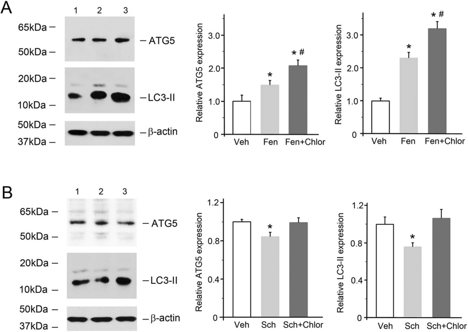
Fig. 4.

D5R-mediated increases in the levels of autophagy-related proteins in D5R-HEK293 cells. A The cells were treated with fenoldopam (Fen, 1.0 μM, 12 h) in the absence or presence of chloroquine (an inhibitor of autophagosome-lysosome fusion/lysosome-mediated proteolysis, 10 μM, 12 h). Immunoblots from one of five independent experiments are shown. Lane 1, vehicle (Veh); lane 2, fenoldopam (Fen); lane 3, fenoldopam plus chloroquine (Fen + Chlor). The β-actin protein level was used to measure the amount of sample loaded in each lane. n = 5/group, *P < 0.05 vs Veh, #P < 0.05 vs Fen. B D5R-HEK293 cells were treated with Sch (Sch23390, 1.0 μM, 12 h) in the absence or presence of chloroquine (Chlor). Immunoblots from one of five independent experiments are shown. Lane 1, Veh; lane 2, Sch; lane 3, Fen + Chlor. The β-actin protein level was used to measure the amount of sample loaded in each lane. n = 5/group, *P < 0.05 vs others
