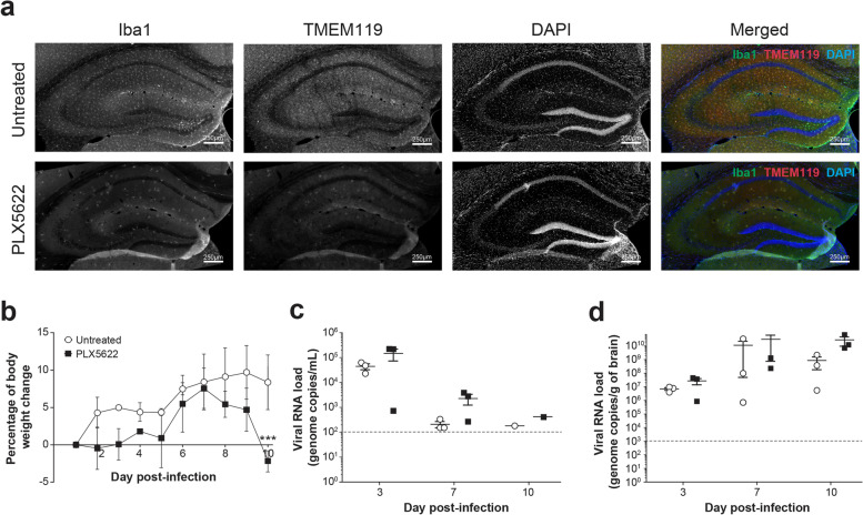
Fig. 6.
Effect of microglia depletion with PLX5622 on ZIKV infection of young adult mouse brain. a In a first experiment, TRIF−/− × IPS-1−/− mice received control or PLX5622 diet ad libitum for 10 days. Mice were sacrificed, and the brain was taken for immunofluorescence analysis. Representative micrographs illustrating the depletion of Iba1+/TMEM119+ microglia after immunofluorescence labeling in the stratum radiatum of untreated (upper images) and PLX5622-treated (lower images) mice. Brain sections were counterstained with DAPI. Scale bars on the picture are equivalent to 250 µm. b–d In a second experiment, TRIF−/− × IPS-1−/− mice received control or PLX5622 diet ad libitum for 10 days. Mice were then infected intravenously with 1 × 10⁵ PFUs of ZIKV strain PRVABC59 (in 100 µL volume), and the respective diets were continued until sacrifice. b Body weight changes of untreated (○) and PLX5622-treated (■) mice after ZIKV challenge. Results are the mean ± SEM of 3 mice per group and per time point. c–d Subsets of mice were sacrificed on day 0 (noninfected), 3, 7, and 10 post-infection, and viral RNA load was determined by reverse transcriptase ddPCR in serum (c) and brain homogenates (d). The dotted lines represent the limits of detection of the reverse transcriptase ddPCR assay. Results are the mean ± SEM of 3 mice per group and per time point. Statistical analyses were performed using a one-way analysis of variance with Tukey’s multiple comparison test. Results that are statistically different between indicated groups are shown as follows: *** P < 0.001

