
Figure 3.
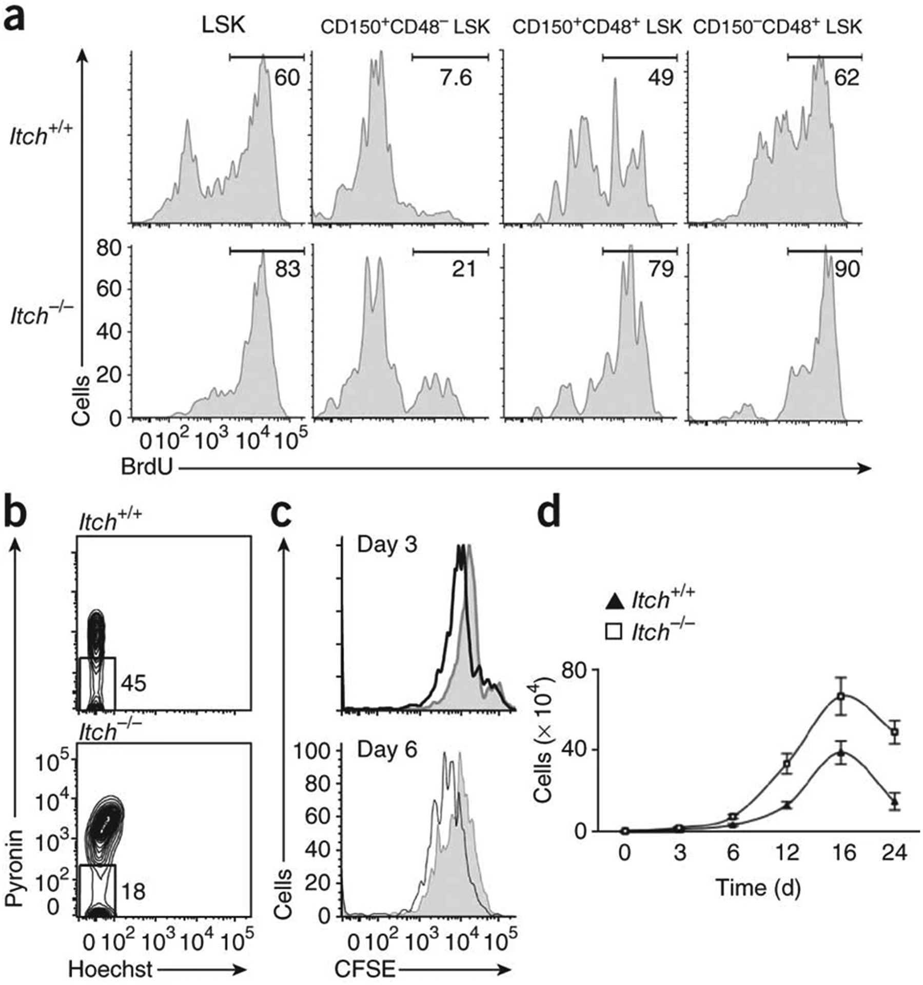
Accelerated proliferation of Itch-mutant HSCs. (a) In vivo proliferative potential of wild-type and Itch−/− LSK subsets, as assessed by BrdU incorporation. Numbers above bracketed lines indicate percentages of proliferating (BrdU+) cells. (b) Ex vivo cell cycle status of wild-type and Itch−/− LSK cells, as assessed by pyronin Y and Hoechst staining. (c) In vitro proliferative potential of wild-type (shaded histograms) and Itch−/− (black lines) CD150+CD48− LSK cells, as assessed by CFSE dilution. (d) Ex vivo population expansion of wild-type and Itch−/− CD150+CD48− LSK cells in response to a cytokine ‘cocktail’. Data are representative of two (a,b) or five (c,d) independent experiments (mean ± s.e.m. of duplicates in d).