
Figure 1.
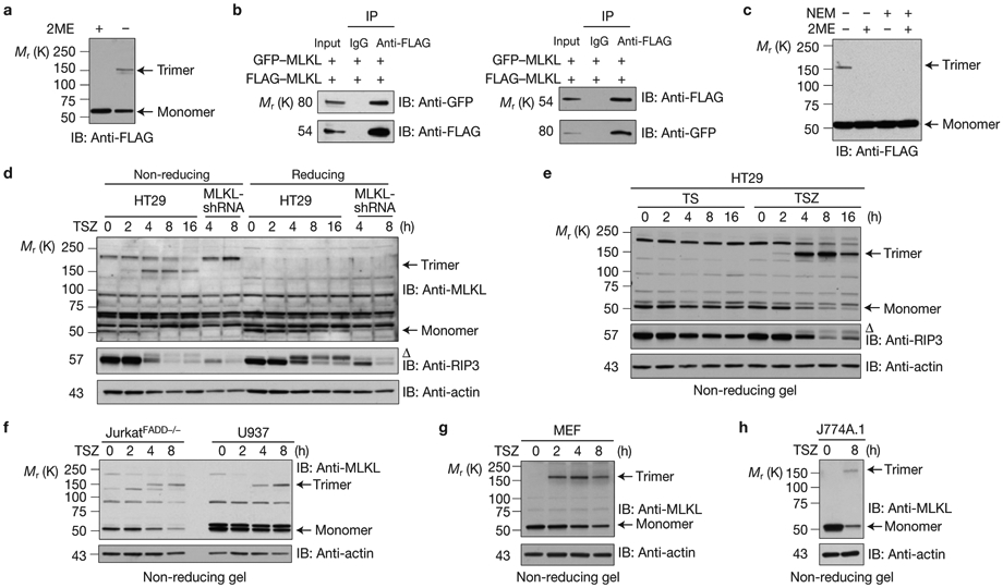
MLKL forms homotrimeric protein on necrosis induction. (a) HEK293 cells were transfected with FLAG–MLKL. The cell lysates were resolved on an SDS–PAGE gel with or without β-mercaptoethanol (2ME) and analysed by immunoblotting with anti-FLAG antibody. (b) HEK293 cells were transfected with GFP–MLKL and FLAG–MLKL as indicated. Cell lysates were immunoprecipitated either with anti-FLAG antibody (left panel) or anti-GFP antibody (right panel) and analysed by immunoblotting with the indicated antibodies. (c) HEK293 cells were transfected with FLAG–MLKL, then lysed with or without 30 mM N-ethylmaleimide (NEM) to block all reactive sulphydryl groups. The cell lysates were resolved with or without 2ME and analysed by immunoblotting with anti-FLAG antibody. (d,e) Control-shRNA or MLKL-shRNA HT29 cells were treated with TSZ (TNF, Smac mimetic and the caspase inhibitor z-VAD-FMK) or TS (TNF and Smac mimetic) as indicated. The cell lysates were resolved either on reducing or non-reducing gel and analysed by immunoblotting with the indicated antibodies. (f–h) JurkatFADD−/−, U937 (f), MEF (g) and J774A.1 (h) cells were treated with TSZ at different time points as indicated. The cell lysates were resolved on non-reducing gel and analysed by immunoblotting with the indicated antibodies. Δ indicates phosphorylated RIP3. All western data are representative of two or three independent experiments. Uncropped images of western blots are shown in Supplementary Fig. 8.