. 2021 Aug 10:211396. doi: 10.1148/radiol.2021211396

Figure 5.

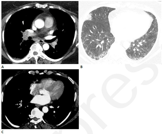
Pulmonary vascular disease after COVID-19 in a 63-year-old woman. (A) CT pulmonary arteriogram with persistent shortness of breath and elevated d-Dimer, seven weeks after onset of infection, shows obstructive thrombus in right interlobar pulmonary artery. (B) CT with lung windows at a lower level shows patchy ground glass opacity, and a focal wedge-shaped consolidative abnormality in the right middle lobe typical for pulmonary infarct. (C) Three months later, the large central thrombus had resolved, but nonocclusive linear webs were present in segmental vessels (arrows), typical for chronic thromboembolic disease.

Pulmonary vascular disease after COVID-19 in a 63-year-old woman. (A) CT pulmonary arteriogram with persistent shortness of breath and elevated d-Dimer, seven weeks after onset of infection, shows obstructive thrombus in right interlobar pulmonary artery. (B) CT with lung windows at a lower level shows patchy ground glass opacity, and a focal wedge-shaped consolidative abnormality in the right middle lobe typical for pulmonary infarct. (C) Three months later, the large central thrombus had resolved, but nonocclusive linear webs were present in segmental vessels (arrows), typical for chronic thromboembolic disease.