Figure 8.

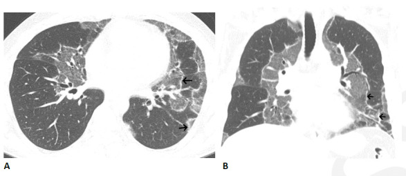
Organizing pneumonia pattern with atoll sign following COVID-19. (A, B) Axial and coronal CT images show multiple areas of sharply demarcated ground glass abnormality with thin peripheral rim (arrows).

Organizing pneumonia pattern with atoll sign following COVID-19. (A, B) Axial and coronal CT images show multiple areas of sharply demarcated ground glass abnormality with thin peripheral rim (arrows).