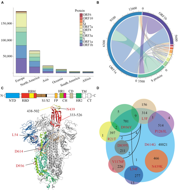
FIGURE 1.
The distribution of Missense Mutations in SARS-CoV-2. A stacked histogram shows all missense mutations of SARS-CoV-2 from different continents of the world. (B) A chord diagram shows the missense mutation shared between different proteins of SARS-CoV-2, and the color scheme is the same as that in (A). (C) A topology and cartoon representation of SARS-CoV-2 homo-trimeric spike (S) glycoprotein (PDB ID: 6VSB). NTD, N-terminal domain; FP, fusion peptide; HR1, heptad repeat 1; CH, central helix; CD, connector domain; HR2, heptad repeat 1; TM, transmembrane region; CT, cytoplasmic tail; S1/S2, protease cleavage sites; Some important mutation sites and residue intervals were marked on the figure. (D) A Venn diagram of main amino acid mutation sites of the S protein, the number of mutations, and co-mutations were shown.

