Figure 7.
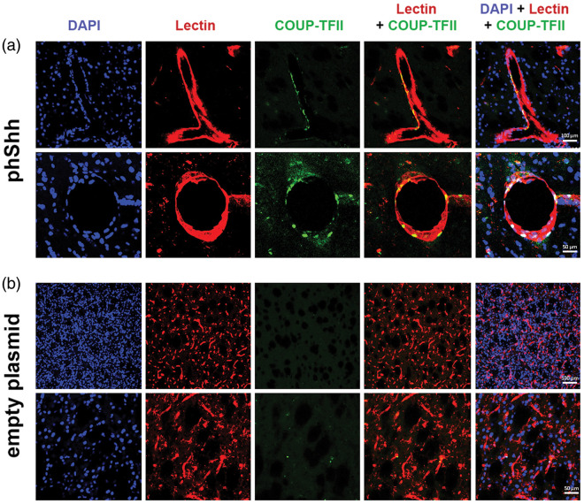
Expression of COUP-TFII in phShh-induced brain neovessels. Sections of brain hemispheres injected with phShh and empty plasmid were stained for DAPI (blue staining) to identify cell nuclei, lectin (red staining) to identify blood vessels, and COUP-TFII (green staining) to identify cells expressing this vascular differentiation marker. (a) COUP-TFII-positive cells were found in the neovessels grown in the brain in response to phShh injection, both at the level of the intimal layer and the vascular wall. (b) COUP-TFII-positive cells were virtually absent in brain hemispheres injected with the empty plasmid.

