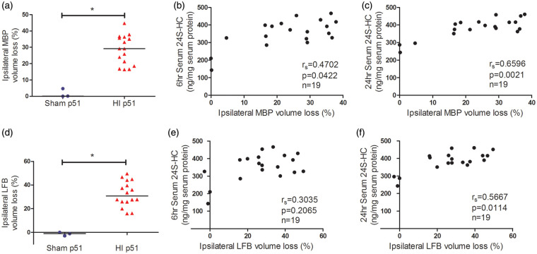
Figure 4.
The serum 24S-HC levels at 24 h after HI were associated with the ipsilateral MBP and LFB volume loss at p51. MBP immunohistochemistry and LFB staining were used to assess brain white matter injury and showed significant volume loss at six weeks (p51) after HI (a and d). The serum 24S-HC concentrations at 24 h (c and f), but not at 6 h (b and e), were highly correlated with the ipsilateral MBP/LFB volume loss. The animal numbers, spearman rs, and p values were listed in the individual panels. For each comparison, the significant level α was specified at 0.05/2 = 0.025 after Bonferroni correction. 24S-HC: 24S-hydroxycholesterol; HI: hypoxia-ischemia; MBP: myelin basic protein; LFB: luxol fast blue. *p < 0.0001.

