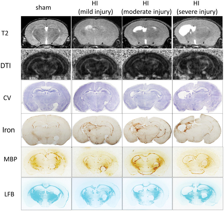
Figure 5.
The representative images of MRI (T2W and DTI-fractional anisotropy map), histology staining (CV and Iron, LFB), and MBP immunohistochemistry. In examples of sham, mild, moderate, and severe HI injury, the injury patterns were similar using the imaging and histology methodology with the damage mainly located in ipsilateral hippocampus, cortex, and thalamus. MBP- and LFB-positive areas are mainly corpus callosum, external capsule, caudate, thalamus, hippocampal fimbria, and internal capsule (note that the circles on the right side of the MBP/LFB sections were punched to label the contralateral hemisphere, not the actual injury). HI: hypoxia-ischemia; MBP: myelin basic protein; LFB: luxol fast blue; CV: cresyl violet; DTI: diffusion tensor imaging.

