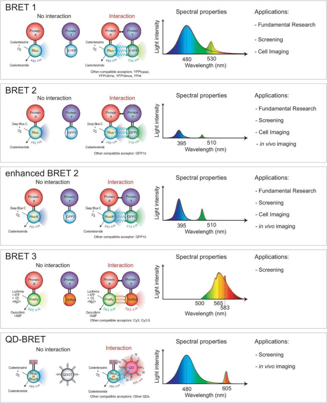
Figure 11.
Schematic representation of different BRET methods. Each method is characterized by different BRET donors and acceptors, spectral properties, and applications. BRET2 uses coelenterazine 400a, which shifts the emission of RLuc to that of GFP and has a larger separation of donor and acceptor emission peaks. Enhanced BRET2 has a light intensity stronger than that of BRET2. BRET3 uses FLuc and DsRed, which has a prolonged emission signal. QD-BRET uses quantum dots and has a distinct separation of donor and acceptor emission peaks. GFP2: green fluorescent protein 2. RLuc8: variant of Renilla luciferase. DsRed: red fluorescent protein. 6-His: polyhistidine tag. QD or QDot: quantum dots. Adapted with permission from ref (60). Copyright 2008 Wiley-VCH Verlag GmbH & Co.

