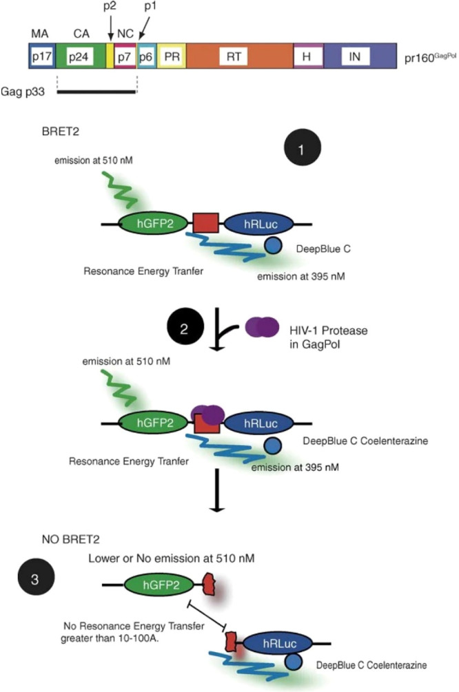
Figure 13.

Schematic representation of BRET-based biosensor to detect HIV-1 protease activity. In the top panel, the proteins in pr160GagPol and protease cleavage sites are shown. In the bottom panel, the process of BRET2 assay is shown: (1) RLuc substrate DeepBlue C is added and interacts with hRLuc, and resonance energy is transferred to hGFP2; (2) HIV-1 protease cleaves its substrate between hGFP2 and hRLuc; (3) resonance energy transfer is low, and there is less light emission from the acceptor molecule. The inset legend on the right panel displays all of the PR cleavage sites and mutant sites tested. pr160GagPol: polyprotein encoded by HIV-1. Gag p33: Gag processing intermediate. p1 and p2: spacer peptides. MA: matrix protein. CA: capsid protein. NC: nucleocapsid protein. PR: protease. RT: reverse transcriptase. H: RNaseH. IN: integrase. hRLuc: humanized sea pansy Renilla reniformis luciferase; donor molecule. hGFP2: humanized green fluorescent protein; acceptor molecule. Adapted with permission from ref (71). Copyright 2005 Elsevier B.V.
