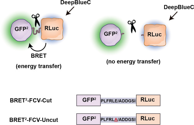
Figure 14.
Schematic representation of wild-type and mutant BRET2-based probes for detection of FCV protease activity. Top: Energy transfer dissipates, and BRET2 signal disappears when GFP2 and RLuc are distanced upon cleavage by the FCV protease. Bottom: Amino acid sequences of the cut and uncut FCV protease cleavage motifs are shown. The mutation is underlined at the bottom uncut construct. FCV protease cleaves peptide bonds right after the glutamic acid (E), indicated by a slash. GFP2: green fluorescent protein 2. RLuc: Renilla luciferase. DeepBlueC: substrate of Rluc.

