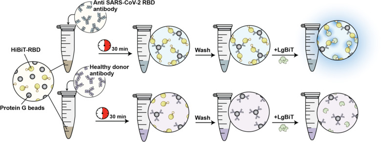
Figure 27.
Schematic representation of the NanoBiT serological assay. RBD fused to HiBiT (HiBiT-RBD) is incubated in the patient sample along with protein G beads. LgBiT detects RBD-binding antibodies in the sample by binding to antibody-bound conjugates and fluorescing (top panel). Adapted with permission from ref (125). Copyright 2021 Azad et al.

