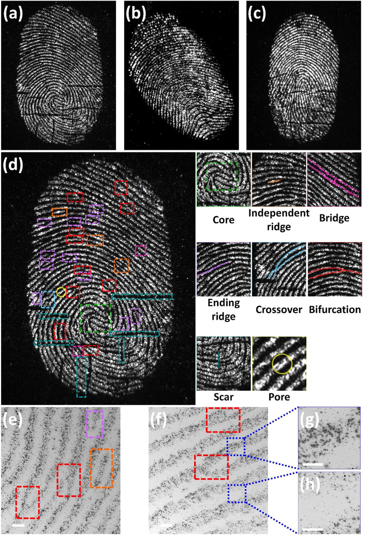
Figure 4.
Fluorescence and SEM images of developed LFPs using FND@PVP. The fluorescence images were obtained with 525 nm excitation, a 30 s exposure, and an Ethidium Bromide (570~640 nm) emission filter. Fingerprint patterns for three donors (a), (b), and (c). (d) High resolution fluorescence image of donor 1’s fingerprint. Unique features are identified inside colored boxes. Magnified fluorescence images of the developed LFP exhibit unique details of the fingerprint such as core (Level 1), independent ridge, bifurcation, ending ridge, crossover, bridge (Level 2), scar, and pores (Level 3). (e) and (f) SEM images show Level 2 features of fingerprint and high interaction between ridges in the LFPs rather than the furrows. (g) and (h) SEM images of ridges and furrows at higher magnification.

