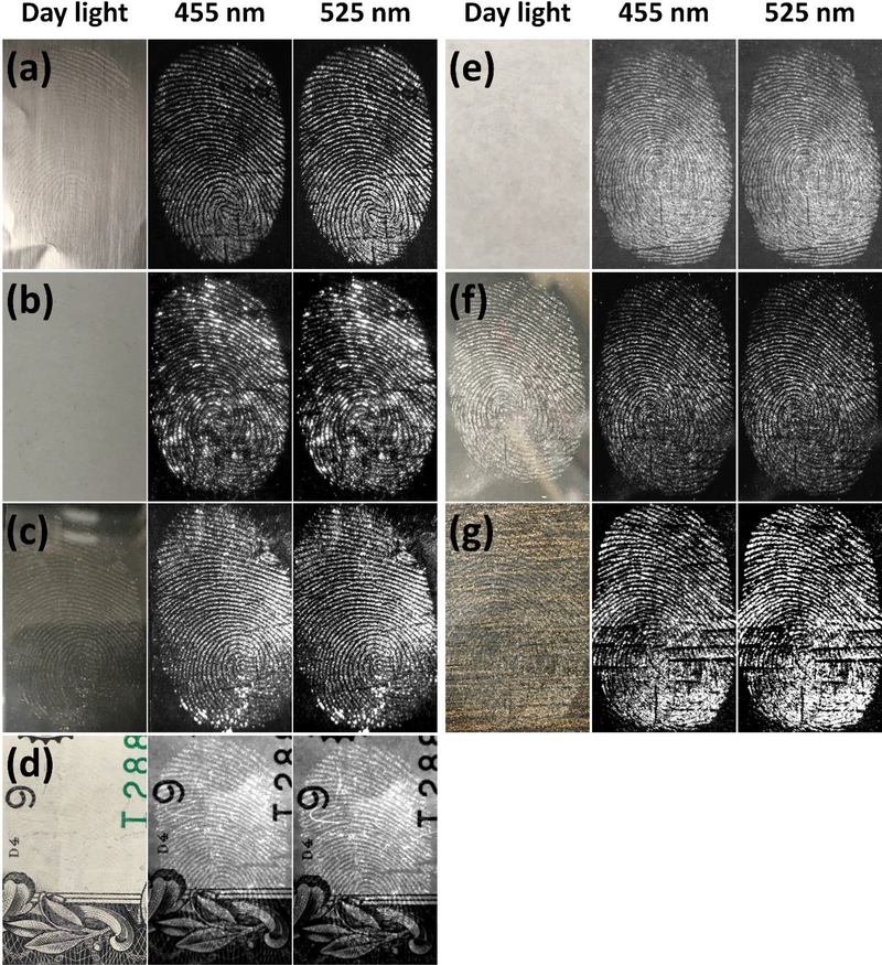
Figure 5.
Bright-field and fluorescence images of FND@PVP treated fingerprints on various surfaces: (a) aluminum foil, (b) ceramic, (c) glass, (d) money (e) paper, (f) plastic petri-dish, and (g) wood. All fluorescence images were obtained with two (455 and 525 nm) excitation wavelengths. Different exposure times were employed for each substrate because the FND@PVP binding differed among substrates. A SYBR Green (515~570 nm) emission filter was employed with 455 nm excitation light, whereas an Ethidium Bromide (570~640 nm) emission filter was employed with 525 nm excitation. The developed LFPs were clearly observed on all surfaces due to strong binding of FND@PVP to finger residues.

