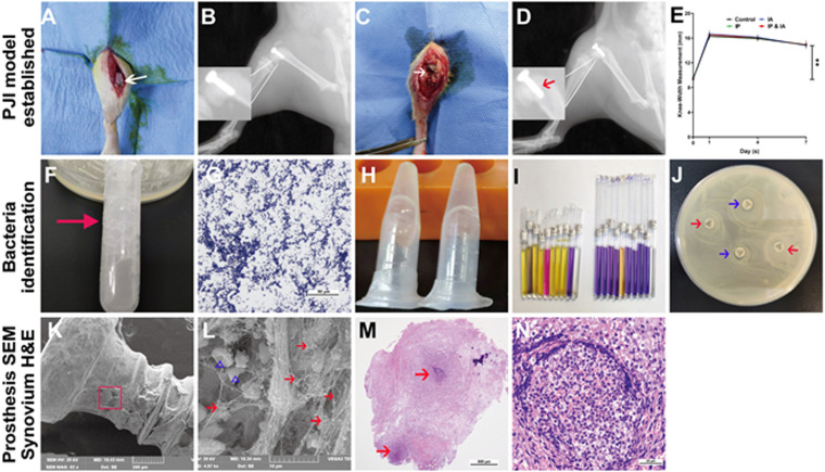
FIG 1.
Establishment and evaluation of the PJI model after knee prosthesis implantation in rats. (A) Artificial prosthesis implanted in the knee joint. (B) Lateral X-ray after surgery. (C) Subcutaneous macroscopic examination of the knee joint on postsurgical day 7. The white arrow indicates subcutaneous and IA abscess or ulcer. (D) Lateral X-ray of the knee on postsurgical day 7. The red arrow indicates prosthesis loosening and suspicious osteolysis around the prosthesis. (E) Changes of the surgical knee width on postsurgical days 1, 4, and 7. (F) Microbial catalase testing (many bubbles appear after the microorganisms contact hydrogen peroxide). (G) Microbial Gram staining (Gram-positive bacteria appear purple blue, indicating positive results). (H) Rapid agglutination testing of fresh rabbit plasma (plasma coagulates in a gelatinous form). (I) Staphylococcus identification kit testing (the corresponding indicator reagent tube changes color, indicating that the microorganism is Staphylococcus aureus). (J) Cefoxitin susceptibility disc diffusion testing in LB agar plates with cultured microorganisms (the antibiotic susceptibility test discs [cefoxitin and norfloxacin] have no inhibition zone, indicating that the bacteria are resistant to cefoxitin and norfloxacin; the red arrows indicate cefoxitin sensitivity test discs, and the blue arrows indicate norfloxacin sensitivity test discs). (K and L) Prosthesis taken from the knee for SEM observations at low magnification (×60) (K) and high magnification (×5,000) (L). The red arrows indicate S. aureus, and the blue triangles indicate red blood cells. (M and N) H&E staining of the surgical knee synovium, at low magnification (×40) (M) and high magnification (×400) (N). The red arrows indicate abscess areas. **, P < 0.01. Significance was evaluated using a two-way ANOVA for the comparison of rat knee widths between different treatment groups.

