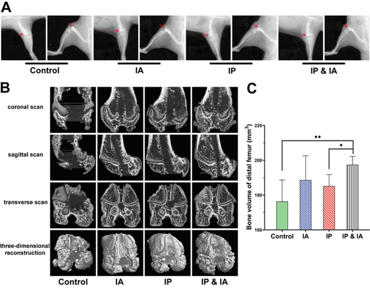
FIG 4.
Radiological evaluation of the knee joint in rats with PJI after debridement and treatment with vancomycin. (A) X-ray of each group on postrevision day 14. (B) Three-dimensional CT scans and bone reconstruction of the distal femur on day 21 (postrevision day 14). (C) Bone volume analysis of the distal femur. Control (no antibiotics), IP injection of vancomycin (88 mg/kg, q12h), IA injection of vancomycin (44 mg/kg, qd), and IP plus IA injection of vancomycin (combined IP treatment at 88 mg/kg, q12h, and IA treatment at 44 mg/kg, qd) were assessed. *, P < 0.05; **, P < 0.01 (n = 6). The red arrows indicate the position of the prosthesis and the destruction of the distal femur in each group. Significance was evaluated using an unpaired one-tailed Mann-Whitney test for the comparison of residual bone volumes in different treatment groups.

