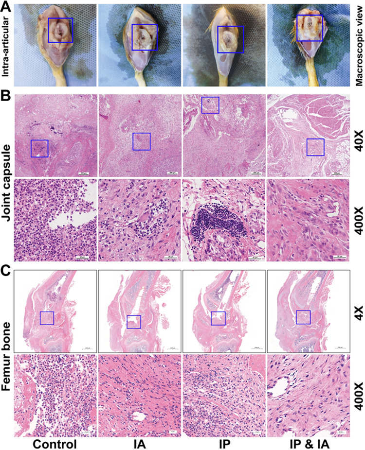
FIG 5.
Macroscopic examination and histopathological assessment of the surrounding knee joint after one-stage revision and treatment with vancomycin in rats with PJI. (A) Macroscopic examination of intraarticular bone and the prosthesis. (B) Pathological H&E staining of joint capsules in each group on day 21. (C) Pathological H&E staining of the knee joint (femur and tibia) in each group on day 21. Control (no antibiotics), IP injection of vancomycin (88 mg/kg, q12h), IA injection of vancomycin (44 mg/kg, qd), and IP and IA injection of vancomycin (combined IP treatment at 88 mg/kg, q12h, and IA treatment at 44 mg/kg, qd) were assessed. The blue boxes in panels A show the position of the prosthesis and the destruction of the cartilage around the prosthesis, the blue boxes in panels B show the area where inflammation is concentrated, and the blue boxes in panels C represent the entrance of the bone tunnel at the femoral trochlea.

