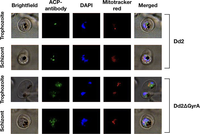
FIG 3.
Loss of apicoplast immunofluorescence in Dd2ΔGyrA clone. The parasite apicoplast was stained with the ACP antibody, which was visualized with the anti-rabbit secondary antibody conjugated to Alexa Fluor 488. Nucleus was stained with DAPI, while mitochondrion was stained with the Mitotracker CMXRos. The apicoplast remained intact in the Dd2 clone, as ACPs were localized to a specific location. However, in the Dd2ΔGyrA clone, ACPs were dispersed throughout the cytoplasm of parasites, indicating loss of the apicoplast structure. Nuclear and mitochondrial genome remained intact in both Dd2 and Dd2ΔGyrA clones.

