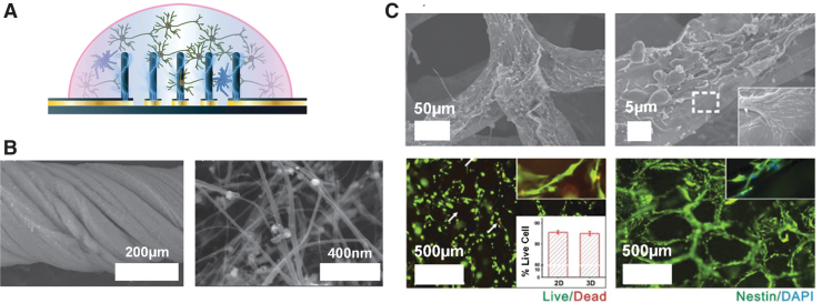
FIG. 2.
(A) PEDOT:PSS pillars were successfully 3D printed using a novel direct-write method, and electrical stimulation was applied to enhance NPC proliferation65; (B) CNT-based electrodes twisted into a rope morphology, which can be effectively utilized for neural stimulation. The graph on the right is the microscopy image of the synthesized CNTs at a higher magnification68; and (C) 3D graphene foam formation utilized as electrically conducting and biocompatible neural scaffolds for NPCs. The top two microscopy images show the structure of the graphene foams in detail and the region outlined with, while dashed line in the right image indicates the interaction between the cell and graphene foam surface. The bottom left image is a cell viability test with NPCs seeded on the graphene foam structure after 5 days of culturing, and the inset is the percentage live cell data (live cells—green, dead cells—red, and the arrows are indicating the dead cells). The bottom right image is a fluorescence image of NPC proliferation on the graphene foam surface (nestin for NPCs—green and DAPI for nuclei—blue).69 3D, three-dimensional; CNTs, carbon nanotubes; NPC, neural precursor cell; PEDOT:PSS, poly(3,4-ethylenedioxythiophene) polystyrene sulfonate. Reproduced with permission. Copyright Wiley (A, B) and Nature (C).

