Figure 2.
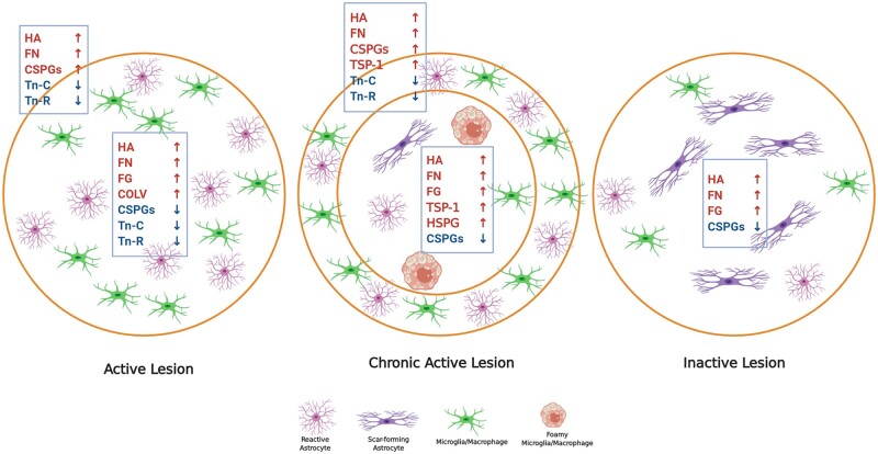
Expression of ECM proteins in different white matter multiple sclerosis lesions. Schematic shows ECM changes in distinct multiple sclerosis lesion types including active, chronic active and inactive lesions. Early demyelinating active lesions consist of hypercellular lesion centre containing reactive astrocytes and immune cells. Chronic active lesions are defined by a hypercellular inflammatory margin and a hypocellular centre with fibrous astrocytes and foamy (myelin-laden) microglia/macrophages. Inactive lesions show minimal signs of inflammation while containing mainly scar forming (fibrous) astrocytes. Each lesion type has a different ECM composition when compared among each other and to normal white matter. Refer to the main text for the capacity of individual ECM components to modulate the activity of immune cells. In active lesions, prominent sources of hyaluronan (HA), fibronectin (FN), CSPGs and Tenascin-C/R (Tn-C/R) appear to be reactive astrocytes while fibrinogen (FG) is deposited by leakage of serum into lesions. Sources of CSPGs are reactive astrocytes and macrophages/microglia although these cells are also removing the deposited CSPGs from the lesion centre as the immune cells move outwards.16 The accumulation of inhibitory ECM such as CSPGs in the lesion edge is thought to be a barrier to incoming progenitor cells such as OPCs that attempt to repair the lesion.3 COLV = collagen V. Images were created using BioRender.

