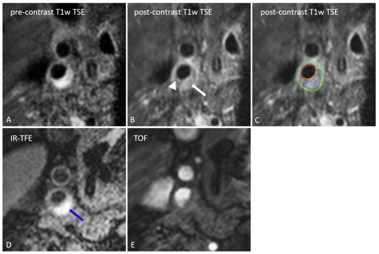
Figure 1.
Transversal MR images of the right internal carotid artery. The black blood pre-contrast image (A) is used to draw the contours of the lumen and outer vessel wall. The lipid-rich necrotic core shows no contrast-enhancement on the post-contrast black-blood T1w quadruple inversion recovery (QIR) turbo spin echo (TSE) image (B) and includes the entire area of hemorrhage (IPH) [IPH: blue, lipid-rich necrotic core: yellow, lumen: red, outer vessel wall: green on (C)]. IPH [blue arrow on (D)] appears as a bright signal on the inversion recovery turbo field echo images (IR-TFE; D). A thin or ruptured fibrous cap (TRFC) can be identified by the interruption of juxtaluminal signal enhancement on the post-contrast T1w image (arrow head). With a contra-indication for contrast injection the T2w image or time of flight (TOF) image (E) can be used for TRFC assessment.

