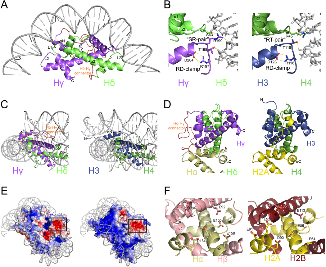
Fig. 3. |. DNA binding and acidic patch conservation.
a Hδ-Hγ “forced dimer”-DNA interface. b, Comparison of interactions between Hδ-Hγ with DNA (left) and human H4 and H3 with DNA (right). c, Close-up view of αN helices and the DNA ends of the Hγ (left) and H3 (right). d, Overview of the C-termini of Hα (left) and H2A (right). e, Representation of electrostatic surface potential of Msv (left) and human (right) nucleosomes. f, Close-up of the acidic patch in Marseillevirus Hβ-Hα (left) and H2B and H2A (right).

