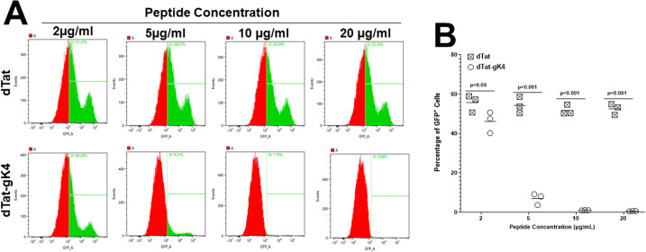
Fig 4. Analysis of infected cells in the presence of gK4 peptide.
(A) Detection of HSV-GFP±cells. Vero cells were infected with 0.1 PFU/cell of HSV-1 expressing GFP in the presence of 2, 5, 10, and 20 μg/ml of dTat-gK4 peptide or control dTat peptide for 1 hr. After 1hr infection, the media was replaced with fresh media containing each peptide. At 24 hr PI, cells were trypsinized, fixed, and the presence of GFP+ cells at each peptide concentration was determined by FACS. B) Quantitation of HSV-GFP± cells. Percentage of GFP+ cells infected as in (A) above were quantitated by FACS. Each point represents the mean ± SEM from four independent experiments.

