FIG 1.
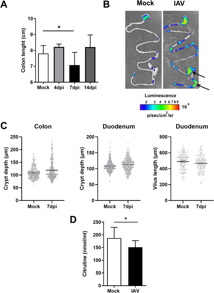
Intestinal inflammation and disorders during IAV infection. Mice were infected or not (mock) with IAV (H3N2). (A) The colon length was measured at 4, 7, and 14 dpi (n = 6 to 13). For the mock control, colons were collected at day 7. (B) Ex vivo bioluminescence imaging was performed on guts collected from naive and IAV-infected (7 dpi) NF-κB–luciferase transgenic mice. Arrows indicate higher NK-κB expression. The scale indicates the average radiance. One representative image of at least 10 mice is shown. (C) Histological analysis of intestinal (colon and duodenum) sections in mock-treated mice and IAV-infected mice (7 dpi). Crypt depths and villus lengths were determined after hematoxylin and eosin coloration (n = 9 to 10/group). (D) Citrulline concentrations in the blood collected from mock-treated mice and IAV-infected mice (7 dpi) (n = 14). Results represent two pooled experiments. Significant differences were determined using the Mann-Whitney U test (C and D) and the Kruskal-Wallis ANOVA test (A). (*, P < 0.05).

