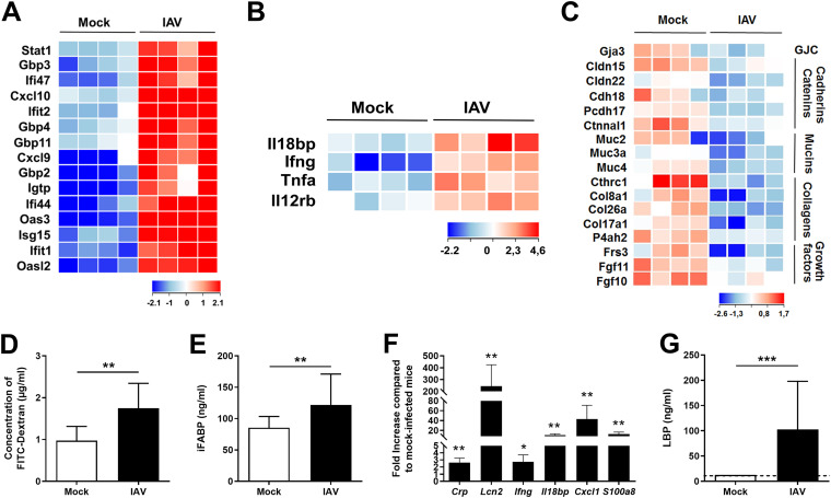
FIG 2.
Altered colonic gene expression and increased gut permeability during IAV infection. (A to C) Transcriptomic analysis of colon samples from mock-treated and IAV-infected mice (7 dpi). Shown are heat maps representing ISGs (A), NF-κB-dependent inflammatory genes (B), and transcriptional expression of components involved in barrier functions and epithelial integrity (GJC, gap junction components) (C) (n = 4/group). (D) Fluorescence intensity quantified in the blood of mice 4 h after FITC-dextran oral administration (n = 8). (E) Intestinal fatty acid-binding protein (iFABP) concentration in the blood (n = 21 to 28). (F) Analysis of gene expression in liver by RT-qPCR (n = 5). (G) LPS-binding protein (LBP) concentration in the blood (n = 11 to 15). (D to G) Results are from two to three pooled experiments (7 dpi). Significant differences were determined using the Mann-Whitney U test (D, E, and G) and the Kruskal-Wallis ANOVA test (F) (*, P < 0.05; **, P < 0.01; ***, P < 0.001).

