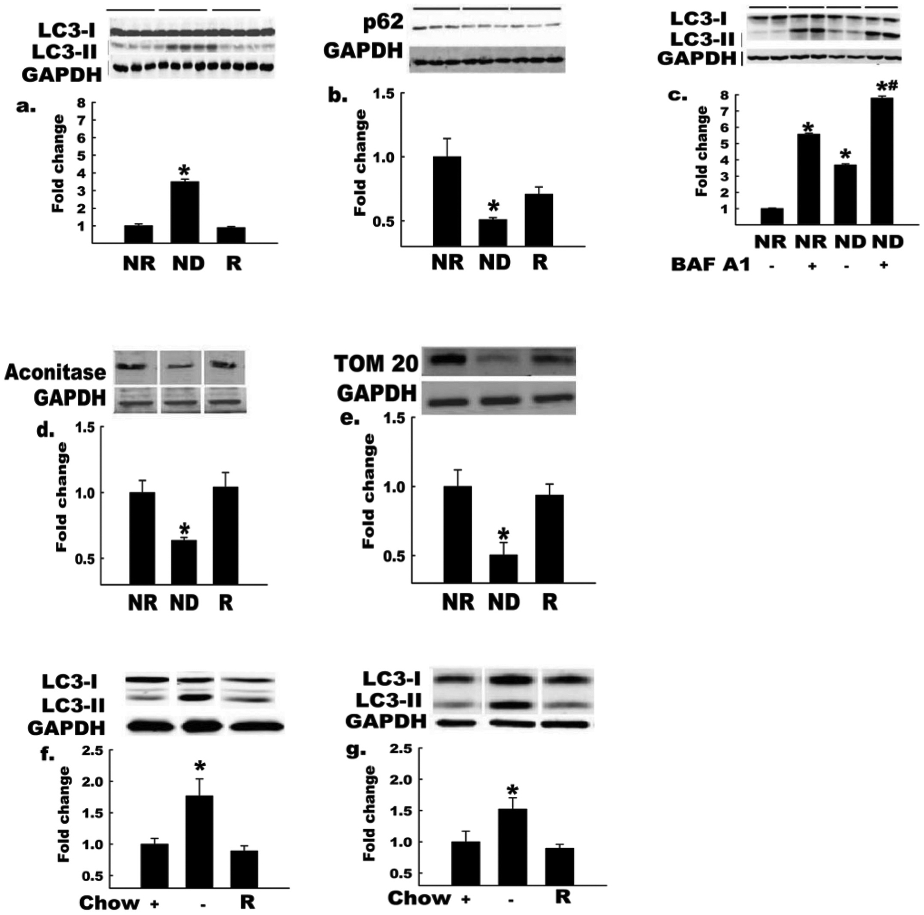
Fig. 1.

Autophagy is increased in endothelial cells and intact arteries by nutrient deprivation. Bovine aortic endothelial cells (BAECs) incubated in nutrient-deplete (ND) or nutrient-replete (NR) medium for 2 h. GAPDH, glyceraldehyde-3-phosphate dehydrogenase. LC3-II:LC3-I ratio (a) increased and p62 protein level (b) decreased (*, p < 0.05) in BAECs incubated in ND vs. NR medium (n = 4). These responses were restored when nutrients were added back to ND medium (R, third histogram). Horizontal bars above the images indicate the respective groups. LC3-II:LC3-I ratio (c) showed a greater increase in BAECs incubated with ND medium in the presence of the lysosomal proton pump inhibitor BAF A1, owing to the expected effect of this treatment to block lysosomal degradation of LC3-II (n = 3 per condition; *, p < 0.05 for BAF A1 effect; #, p < 0.05 for −AA effect). Protein levels of the mitochondrial markers m-aconitase and TOM 20 (d, e) decreased in cells incubated in ND vs. NR medium (n = 4, p < 0.05), suggesting increased mitochondrial turnover; in both cases (d, e), responses were reversed when nutrients were restored to ND medium (R). LC3-II:LC3-I increased (*, p < 0.05) in hearts (f) and arteries (g) from in vivo fasted (14 h) vs. fed mice, and these responses were reversed by 1 h re-feeding (R, n = 3 per group). Histograms (below) represent the mean ± SE of densitometry (above). *, p < 0.05 for ND medium (d,e) or fasting (Chow −) (f, g) effect. For Figs. 1a–1e, each n refers to one 10 cm Petri dish. For Figs. 1f and 1g, each n refers to one mouse.
